Hysteroscopy

Hysteroscopy is a minimally invasive procedure that allows a gynaecologist to examine the inside of the uterus (womb) using a hysteroscope, which is a thin, lighted telescope-like instrument.

This procedure can be both diagnostic and operative, making it a valuable tool in the diagnosis and treatment of various uterine conditions.

Hysteroscopy can be performed in an outpatient setting, a clinic, or a hospital, depending on the complexity of the case.

Our team of gynaecologists routinely perform hysteroscopy in-rooms at our clinic, to diagnose a range of conditions. If you have a condition requiring colposcopy, please get in touch to book an appointment.

Applications of Hysteroscopy

Hysteroscopy is used for both diagnostic and therapeutic purposes. It allows gynaecologists to directly view the uterine cavity, identify abnormalities, and perform surgical procedures if necessary. Common applications include:
  • Diagnostic Hysteroscopy: Used to investigate abnormal uterine bleeding, recurrent miscarriages, or infertility issues.
  • Operative Hysteroscopy: Involves the treatment of uterine conditions such as polyps, fibroids, adhesions (Asherman’s syndrome), and septa.

Conditions Diagnosed and Treated by Hysteroscopy

Uterine polyps are benign (non-cancerous) growths attached to the inner wall of the uterus and extending into the uterine cavity.

  • Causes: Hormonal imbalances, particularly an excess of oestrogen.
  • Symptoms: Irregular menstrual bleeding, bleeding between periods, excessive menstrual bleeding, and postmenopausal bleeding.
  • Risk Factors: Age (more common in women in their 40s and 50s), obesity, hypertension, and hormone replacement therapy.
  • Treatment Options: Removal during operative hysteroscopy.

Uterine fibroids (leiomyomas) are non-cancerous growths of the uterus that often appear during childbearing years.

  • Causes: Genetic changes, hormonal factors (oestrogen and progesterone), and growth factors.
  • Symptoms: Heavy menstrual bleeding, prolonged periods, pelvic pain and pressure, frequent urination, constipation, and backache.
  • Risk Factors: Family history, African-Australian ethnicity, obesity, and early menarche.
  • Treatment Options: Removal using hysteroscopic myomectomy.

Endometrial hyperplasia is a condition characterised by the thickening of the uterine lining (endometrium), often caused by excess oestrogen without sufficient progesterone.

  • Causes: Hormonal imbalances, particularly an excess of oestrogen.
  • Symptoms: Heavy menstrual bleeding, bleeding between periods, and postmenopausal bleeding.
  • Risk Factors: Obesity, polycystic ovary syndrome (PCOS), hormone replacement therapy, and tamoxifen use.
  • Treatment Options: Biopsy during hysteroscopy for diagnosis, followed by medical or surgical management based on the findings.

Asherman’s syndrome involves the formation of scar tissue (adhesions) within the uterine cavity, often as a result of surgical procedures like dilation and curettage (D&C).

  • Causes: Uterine surgery, infections, or trauma.
  • Symptoms: Reduced menstrual flow, missed periods, pelvic pain, and infertility.
  • Risk Factors: Previous uterine surgery, infections, and trauma to the uterine lining.
  • Treatment Options: Adhesiolysis (removal of adhesions) using hysteroscopy.

A uterine septum is a congenital condition where a band of tissue (septum) divides the uterine cavity, potentially leading to fertility issues and recurrent miscarriages.

  • Causes: Congenital abnormality during fetal development.
  • Symptoms: Recurrent miscarriages, preterm labour, and infertility.
  • Risk Factors: Family history of uterine abnormalities.
  • Treatment Options: Surgical resection of the septum using hysteroscopy.

The Hysteroscopy Procedure

  • Consultation: Discuss medical history, medications, and allergies with your gynaecologist.
  • Timing: The procedure is usually scheduled after menstruation and before ovulation to ensure the uterine lining is thin.
  • Medication: You may be advised to take pain relief or anti-inflammatory medication before the procedure.
  • Anaesthesia: Depending on the complexity, local, regional, or general anaesthesia may be used.
  • Insertion: A speculum is inserted into the vagina to allow access to the cervix, which is then dilated to insert the hysteroscope.
  • Examination: The hysteroscope is gently guided into the uterine cavity, and saline solution is used to expand the uterus for better visibility.
  • Intervention: If operative hysteroscopy is performed, surgical instruments are inserted through the hysteroscope to treat the identified condition.
  • Recovery: Most women can go home the same day. Mild cramping or spotting may occur.
  • Activity: Avoid heavy lifting, strenuous exercise, and intercourse for a few days as advised by your gynaecologist.
  • Follow-Up: A follow-up appointment may be scheduled to discuss results and further treatment if necessary.

Risks and Complications

While hysteroscopy is generally safe, potential risks and complications include:
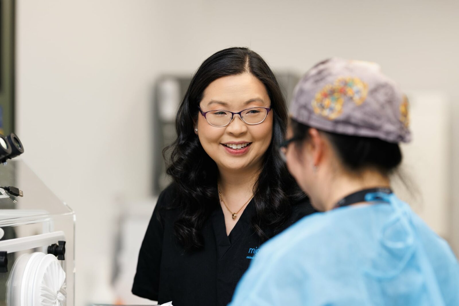
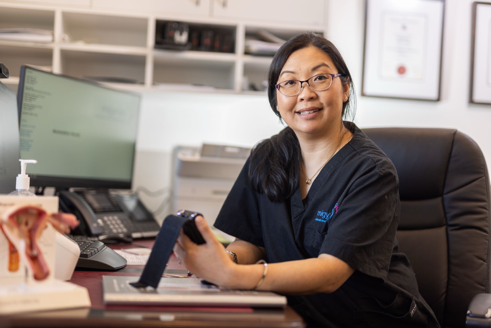
  • Infection: Though rare, there is a risk of uterine or pelvic infection.
  • Bleeding: Some bleeding or spotting is normal, but heavy bleeding should be reported.
  • Uterine Perforation: Accidental perforation of the uterus is rare but can occur.
  • Fluid Overload: During the procedure, the fluid used to expand the uterus can sometimes be absorbed into the bloodstream, causing complications.

Get in touch with our friendly team.

We are a general gynaecology clinic based in Melbourne, dedicated to the latest Minimally Invasive (MI) gynaecological diagnostic and surgical techniques. We are leaders in laparoscopic and cutting-edge robotic “keyhole” surgery.

If you have a question about a condition or treatment, or would like to book an appointment, please get in touch.